
https://www.youtube.com/@psyopchannel13 https://gab.com/boxoffrogs https://rumble.com/c/c-2352810 https://odysee.com/@HNiCC:1
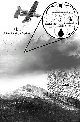
Except it's not silver iodine... it's aluminum, strontium, and beryllium... all much cheaper and much more toxic.
https://www.youtube.com/@psyopchannel13 https://gab.com/boxoffrogs https://rumble.com/c/c-2352810 https://odysee.com/@HNiCC:1
Weather made here...


Silver nanoparticles produce a stronger plasmon resonance than gold nanoparticles it can be due to the large reflectivity from the metal surface. TEM images indicate that change in the size of nanoparticles with increasing the laser fluence is in good agreement with the Mie theory.
Magnetic hyperthermia aims to produce the local heating by a magnetically-mediated heating of low-frequency electromagnetic waves, through the power absorption by magnetic nanoparticles. This technique is one of the most important approaches to induce the local heating by low electromagnetic radiation.
In addition, an applied magnetic field can change the magnetic moment of the object itself; for example by magnetizing it. This phenomenon is known as magnetism. An applied magnetic field can flip the magnetic dipoles that make up the material causing both paramagnetism and ferromagnetism. Additionally, the magnetic field can affect the currents that create the magnetic fields (such as the atomic orbits) which causes diamagnetism.
The vicinity of metallic nanostructures that provides intense optical fields is termed as “hot spot”. Any molecule in close proximity to these hot spots will give rise to an increased surface-enhanced Raman spectroscopic (SERS) signal. We have synthesized Ag core−Au shell (core−shell) nanoparticles (NP) with nanopores, which act as hot spots in the SERS measurements. We have demonstrated a large enhancement in SERS studies of various molecules using core−shell NP with hot spots, which is better than using silver nanoparticles. The core−shell NP with hot spots can be used for ultratrace analysis of important biomolecules such as histone acetyltranferase p300, a human transcriptional coactivator protein. The core−shell NP does not change the biomolecule's physical and chemical property upon adsorption, which makes it biocompatible. The core−shell NP carrying hot spots with high SERS enhancement would be ideally suited for in vivo studies of biological systems.
Plasmonic optical trapping is based on the enhanced electromagnetic field by SPs and have two typical modes: one mode is using the focused laser to trap plasmonic nanoparticles while the other mode is using the plasmonic nanostructure to trap dielectric nanoparticles.
This thesis focuses on cavity optomechanics with an optically levitated dielectric ob- ject.
https://d-nb.info/1045023477/34
Optical resonators with whispering-gallery modes-part II: applications | IEEE Journals & Magazine | IEEE Xplore
We review photonic applications of dielectric whispering-gallery mode (WGM) resonators-tracing the growth of the technology from experiments with levitating droplets of aerosols to ultrahigh-Q solid state crystalline and integrated on-chip microresonators.
https://ieeexplore.ieee.org/document/1588879

Optical nonlinearity involved switching draws an important consideration in nonlinear optical studies. Based on that, we explored nonlinear absorption processes in silver nanoparticles synthesized by liquid phase laser ablation technique employing a second harmonic wavelength (532 nm) of Q switched Nd:YAG laser pulses with 7 ns pulse width and 10 Hz repetition rates. The typical surface plasmon resonance induced absorption (~418 nm) confirmed the formation of Ag NPs. The Z-scan technique was used to study the nonlinear optical processes, employing the same laser system used for ablation. Our study reveals that there is an occurrence of a saturable to reverse saturable absorption switching activity in the Ag nanoparticles, which is strongly on-axis input intensity dependent as well. The closed aperture Z-scan analysis revealed the self-defocusing nature of the sample.