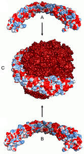
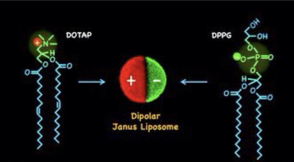
Virosomes are reconstituted liposomes containing viral (typically influenza virus) proteins in the liposomal membrane and, optionally, with additional antigens incorporated in the liposomal membrane or attached to the membrane.
From: Vaccines (Sixth Edition), 2013
Applying ultrasound waves in the region where magnetic erythrocytes accumulate can induce vehicle destruction and the release of a drug at the organ or tissue level [24], [25].
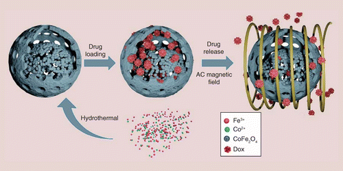
The magnetic erythrocytes, resulting from the co-encapsulation of the drugs with some ferrofluids such as cobalt–ferrite and magnetite, have been reported to direct the encapsulated drug predominantly to the desired sites of the body by means of an external magnetic field [21]–[23].
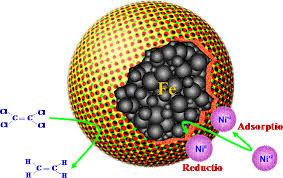
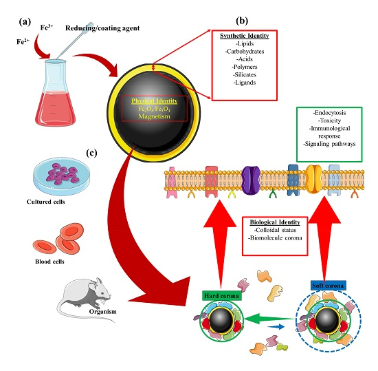
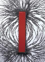
Magnetic nanoparticles are a class of nanoparticle that can be manipulated using magnetic fields. Such particles commonly consist of two components, a magnetic material, often iron, nickel and cobalt, and a chemical component that has functionality. While nanoparticles are smaller than 1 micrometer in diameter (typically 1–100 nanometers), the larger microbeads are 0.5–500 micrometer in diameter. Magnetic nanoparticle clusters that are composed of a number of individual magnetic nanoparticles are known as magnetic nanobeads with a diameter of 50–200 nanometers.
[1][2] Magnetic nanoparticle clusters are a basis for their further magnetic assembly into magnetic nanochains.
[3] The magnetic nanoparticles have been the focus of much research recently because they possess attractive properties which could see potential use in catalysis including nanomaterial-based catalysts,[4] biomedicine [5] and tissue specific targeting,[6] magnetically tunable colloidal photonic crystals,[7] microfluidics,[8] magnetic resonance imaging,[9] magnetic particle imaging,[10] data storage,[11][12] environmental remediation,[13] nanofluids,[14][15] optical filters,[16] defect sensor,[17] magnetic cooling[18][19] and cation sensors.
Ferrite nanoparticles or iron oxide nanoparticles (iron oxides in crystal structure of maghemite or magnetite) are the most explored magnetic nanoparticles up to date. Once the ferrite particles become smaller than 128 nm[22] they become superparamagnetic which prevents self agglomeration since they exhibit their magnetic behavior only when an external magnetic field is applied.
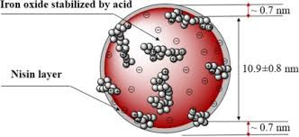
The magnetic moment of ferrite nanoparticles can be greatly increased by controlled clustering of a number of individual superparamagnetic nanoparticles into superparamagnetic nanoparticle clusters, namely magnetic nanobeads.[1] With the external magnetic field switched off, the remanence falls back to zero. Just like non-magnetic oxide nanoparticles, the surface of ferrite nanoparticles is often modified by surfactants, silica,[1] silicones or phosphoric acid derivatives to increase their stability in solution.[23]
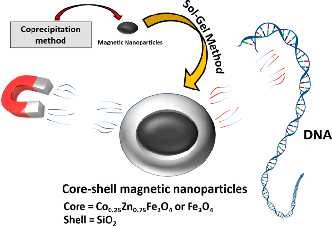
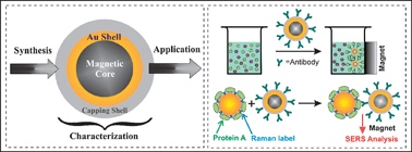
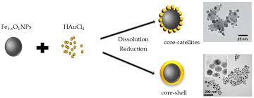
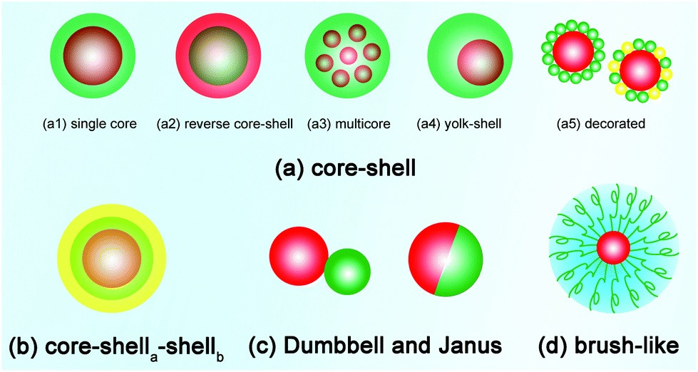
A magnetic core-shell nanoparticle, as the name implies, is a type of nanoparticle having a core or inner magnetic material and an outer shell composed of coating material. Varieties of core-shell nanoparticles have been prepared with diverse composition in close interaction and distinct use. 5 Apr 2017
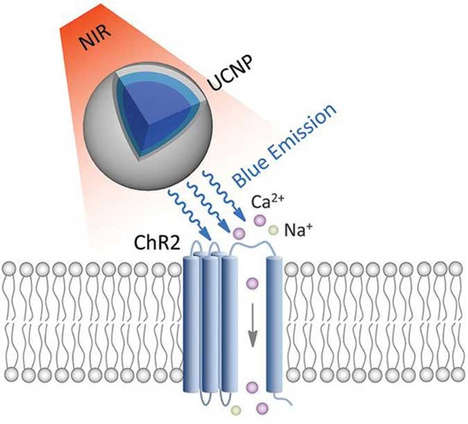
Optogenetics – the use of optically-activated proteins to control neuronal function – is a recent development in neuroscience methodology. Optogenetic techniques provide a means of activating or inhibiting distinct populations of neurons with an unprecedented degree of spatial, temporal, and neurochemical precision. Channelrhodopsin-2 (ChR2), an algal protein from Chlamydomonas reinhardtii, is a light-activated cation channel capable of inducing depolarization and action potentials in neurons.
Limiting Ostwald ripening is fundamental in modern technology for the solution synthesis of quantum dots...


