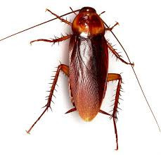
Our current understanding of insect phototransduction is based on a small number of species, but insects occupy many different visual environments. We created the retinal transcriptome of a nocturnal insect, the cockroach, Periplaneta americana to identify proteins involved in the earliest stages of compound eye phototransduction, and test the hypothesis that different visual environments are reflected in different molecular contributions to function.
We assembled five novel mRNAs: two green opsins, one UV opsin, and one each TRP and TRPL ion channel homologs. One green opsin mRNA (pGO1) was 100–1000 times more abundant than the other opsins (pGO2 and pUVO), while pTRPL mRNA was 10 times more abundant than pTRP, estimated by transcriptome analysis or quantitative PCR (qPCR). Electroretinograms were used to record photoreceptor responses. Gene-specific in vivo RNA interference (RNAi) was achieved by injecting long (596–708 bp) double-stranded RNA into head hemolymph, and verified by qPCR.
RNAi of the most abundant green opsin reduced both green opsins by more than 97% without affecting UV opsin, and gave a maximal reduction of 75% in ERG amplitude 7 days after injection that persisted for at least 19 days.
RNAi of pTRP and pTRPL genes each specifically reduced the corresponding mRNA by 90%. Electroretinogram (ERG) reduction by pTRPL RNAi was slower than for opsin, reaching 75% attenuation by 21 days, without recovery at 29 days. pTRP RNAi attenuated ERG much less; only 30% after 21 days. Combined pTRP plus pTRPL RNAi gave only weak evidence of any cooperative interactions. We conclude that silencing retinal genes by in vivo RNAi using long dsRNA is effective, that visible light transduction in Periplaneta is dominated by pGO1, and that pTRPL plays a major role in cockroach phototransduction.
Animal phototransduction proceeds through light absorption by rhodopsins (opsins linked to retinal molecules), which activate ion channels via G-protein coupled second messenger pathways (Fain et al., 2010). Insect and other arthropod compound eyes contain the opsin molecules in microvilli of photoreceptor cells. Rich genetic and molecular tools have made Drosophila compound eyes the best-understood model of insect phototransduction (Hardie and Postma, 2008).
In Drosophila, photon absorption by rhodopsin causes photoisomerization to metarhodopsin, which activates a heterotrimeric Gq-protein, initiating a cascade leading to activation of IP3 and diacylglycerol. Linkages from this cascade to opening of transient receptor potential (dTRP) and TRP-like (dTRPL) ion channels that carry the receptor current are still debated, and both chemical (Chyb et al., 1999; Huang et al., 2010) and mechanical (Hardie and Franze, 2012) intermediate steps have been proposed
In Drosophila, dTRP and dTRPL channels are thought to carry approximately equal parts of light-activated current under physiological conditions (Reuss et al., 1997).
Although major features of Drosophila phototransduction may apply to other insect species, there are probably many variations to accommodate the different visual requirements of this large and diverse group of animals. However, the study of such mechanisms has been hindered by the lack of powerful methods that can be used in Drosophila, including deletion/inactivation mutants and targeted mutations.
Here we used the American cockroach, Periplaneta americana, which has a very different lifestyle to Drosophila, including mainly terrestrial, secluded habitats, reliance on chemical and mechanical information via prominent antennae and cerci, and preference for dark or crepuscular visual environments (Cameron, 1961).
Evidence already exists that the anatomy and physiology of Periplaneta compound eyes are adapted to dim light (Heimonen et al., 2006, 2012), and recent electrophysiological data suggested that these differences include a larger role for TRPL than TRP channels (Immonen et al., 2014).
To explore phototransduction mechanisms we created a transcriptome of Periplaneta retina and assembled mRNA sequences for opsins, TRP and TRPL genes (French, 2012). To investigate the roles of each protein, we used in vivo RNA interference (RNAi) based gene silencing to suppress translation of these genes by injecting double stranded RNA (dsRNA) into the head hemolymph.
Changes in photoreceptor function were measured by an electroretinogram (ERG) assay and the quantity of targeted mRNA measured by qPCR. Our data indicate that Periplaneta retina contains three opsins (pGO1, pGO2, and pUVO), with one of the green opsins, pGO1, dominating vision of visible light. Periplaneta pTRPL mRNA was 10-fold more abundant than pTRP, and RNAi of pTRPL was much more effective in reducing ERG, supporting a more important role for pTRPL than pTRP in Periplaneta phototransduction.
All animal procedures followed protocols approved by the Dalhousie University Committee on Laboratory Animals. Cockroaches, Periplaneta americana, were raised and maintained in the laboratory at a temperature of 22 ± 2°C under a 13 h light/11 h dark cycle.. .
Opsin proteins are fundamental components of animal vision whose structure largely determines the sensitivity of visual pigments to different wavelengths of light. Surprisingly little is known about opsin evolution in beetles, even though they are the most species rich animal group on Earth and exhibit considerable variation in visual system sensitivities. We reveal the patterns of opsin evolution across 62 beetle species and relatives.
Our results show that the major insect opsin class (SW) that typically confers sensitivity to “blue” wavelengths was lost ~300 million years ago, before the origin of modern beetles. We propose that UV and LW opsin gene duplications have restored the potential for trichromacy (three separate channels for colour vision) in beetles up to 12 times and more specifically, duplications within the UV opsin class have likely led to the restoration of “blue” sensitivity up to 10 times.
This finding reveals unexpected plasticity within the insect visual system and highlights its remarkable ability to evolve and adapt to the available light and visual cues present in the environment.
Etymology
tri- + Ancient Greek χρῶμα (khrôma, “color”).
Noun
trichromacy (countable and uncountable, plural trichromacies)
The quality of having three independent channels for conveying color information in the eye.
Trichromacy or trichromatism is the possessing of three independent channels for conveying color information, derived from the three different types of cone cells in the eye. Organisms with trichromacy are called trichromats.
The normal explanation of trichromacy is that the organism's retina contains three types of color receptors (called cone cells in vertebrates) with different absorption spectra. In actuality the number of such receptor types may be greater than three, since different types may be active at different light intensities. In vertebrates with three types of cone cells, at low light intensities the rod cells may contribute to color vision.
Humans and some other mammals have evolved trichromacy based partly on pigments inherited from early vertebrates. In fish and birds, for example, four pigments are used for vision. These extra cone receptor visual pigments detect energy of other wavelengths, sometimes including ultraviolet.
Trichromatic color vision is the ability of humans and some other animals to see different colors, mediated by interactions among three types of color-sensing cone cells.
The trichromatic color theory began in the 18th century, when Thomas Young proposed that color vision was a result of three different photoreceptor cells. From the middle of the 19th century, in his Treatise on Physiological Optics, Hermann von Helmholtz later expanded on Young's ideas using color-matching experiments which showed that people with normal vision needed three wavelengths to create the normal range of colors. Physiological evidence for trichromatic theory was later given by Gunnar Svaetichin (1956).
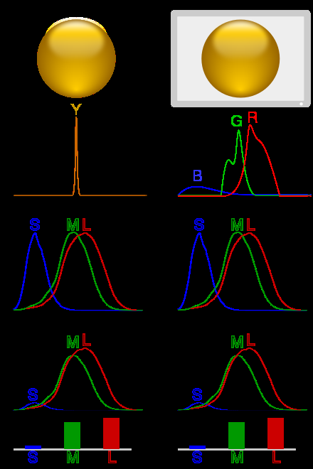
Illustration of colour metamerism:
In column 1, a ball is illuminated by monochromatic light. Multiplying the spectrum by the cones' spectral sensitivity curves gives the response for each cone type.
In column 2, metamerism is used to simulate the scene with blue, green and red LEDs, giving a similar response.
Each of the three types of cones in the retina of the eye contains a different type of photosensitive pigment, which is composed of a transmembrane protein called opsin and a light-sensitive molecule called 11-cis retinal.
Each different pigment is especially sensitive to a certain wavelength of light (that is, the pigment is most likely to produce a cellular response when it is hit by a photon with the specific wavelength to which that pigment is most sensitive). The three types of cones are L, M, and S, which have pigments that respond best to light of long (especially 560 nm), medium (530 nm), and short (420 nm) wavelengths respectively.
Optogenetics: ↑ A technique that uses a combination of light and genetic engineering to control the activity of a cell. ... Opsins: ↑ Proteins that respond to a specific type of light (for example, ChR2 only responds to blue light). In neuroscience, these proteins are used to control neuron activity.
Since the likelihood of response of a given cone varies not only with the wavelength of the light that hits it but also with its intensity, the brain would not be able to discriminate different colors if it had input from only one type of cone. Thus, interaction between at least two types of cone is necessary to produce the ability to perceive color. With at least two types of cones, the brain can compare the signals from each type and determine both the intensity and color of the light.
Optogenetics is a technique used for the study of neural circuits in the brain. It is a branch of biotechnology that combines genetics and optical techniques to conceive and control a specific neural circuit in a living human brain.
Even though optogenetics is a relatively new neuromodulation tool whose various implications have not yet been scrutinized, it has already been approved for its first clinical trials in humans. 17 Oct 2020
scrutinize (third-person singular simple present scrutinizes, present participle scrutinizing, simple past and past participle scrutinized)
(transitive) To examine something with great care or detail, as to look for hidden or obscure flaws.
to scrutinize the conduct or motives of individuals
(transitive) To audit accounts etc in order to verify them.
To shed light on something...
Viral vectors are tools commonly used by molecular biologists to deliver genetic material into cells. This process can be performed inside a living organism or in cell culture. Viruses have evolved specialized molecular mechanisms to efficiently transport their genomes inside the cells they infect.
"enhanced delivery injection of the viral optogenetic vector"
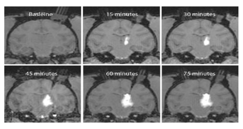
Convection enhanced delivery, or CED, of optogenetic viral vectors in non human primates will enable researchers to manipulate neural activity at a large scale to understand complex neural computations and behaviors. With CED we can achieve high optogenetic expression across large regions of the primate brain with only a few injections in a short amount of time compared to traditional methods.
Magnetic resonance or MR compatible chamber implantation is performed using standard practice in non human primate surgeries, the procedure is described in detail in the text protocol and outlined briefly in this video.
Following animal sedation, positioning, and initial incision as described in the text protocol..
mRNA, or messenger RNA vaccines, borrow a mechanism that all of your cells use to make all the proteins in your body. It helps translate the message in your DNA's genetic code into proteins. 2 days ago
Neuropsin is a protein that in humans is encoded by the OPN5 gene. It is a photoreceptor protein sensitive to ultraviolet (UV) light. ... Neuropsin is bistable at 0 °C and activates a UV-sensitive, heterotrimeric G protein Gi-mediated pathway in mammalian and avian tissues.
Opsins are members of the G protein-coupled receptor superfamily. Human neuropsin is expressed in the eye, brain, testes, and spinal cord. Neuropsin belongs to the seven-exon subfamily of mammalian opsin genes that includes peropsin (RRH) and retinal G protein coupled receptor (RGR). Neuropsin has different isoforms created by alternative splicing
When reconstituted with 11-cis-retinal, mouse and human neuropsins absorb maximally at 380 nm. When illuminated these neuropsins are converted into blue-absorbing photoproducts (470 nm), which are stable in the dark. The photoproducts are converted back to the UV-absorbing form, when they are illuminated with orange light (> 520 nm).
Neuropsin and its orthologs have been found experimentally in a small number of animals, among them human, house mouse (Mus musculus),[5] chicken (Gallus gallus domesticus),[9][12] the Japanese quail (Coturnix japonica),[13] the European brittle star Amphiura filiformis (related to starfish),[14] the tardigrade water bear (Hypsibius dujardini),[15] and the tadpole of Xenopus laevis.[16]
Searches of publicly available databases of genetic sequences have found putative neuropsin orthologs in both major branches of Bilateria: protostomes and deuterostomes. Among protostomes, putative neuropsins have been found in the molluscs owl limpet (Lottia gigantea) (a species of sea snail) and Pacific oyster (Crassostrea gigas), in the water flea (Daphnia pulex) (an arthropod), and in the annelid worm Capitella teleta.
Opsins are a group of proteins made light-sensitive via the chromophore retinal (or a variant) found in photoreceptor cells of the retina. Five classical groups of opsins are involved in vision, mediating the conversion of a photon of light into an electrochemical signal, the first step in the visual transduction cascade. Another opsin found in the mammalian retina, melanopsin, is involved in circadian rhythms and pupillary reflex but not in vision.
Opsins can be classified several ways, including function (vision, phototaxis, photoperiodism, etc.), type of chromophore (retinal, flavine, bilin), molecular structure (tertiary, quaternary), signal output (phosphorylation, reduction, oxidation), etc.
Opsin proteins covalently bind to a vitamin A-based retinaldehyde chromophore through a Schiff base linkage to a lysine residue in the seventh transmembrane alpha helix. In vertebrates, the chromophore is either 11-cis-retinal (A1) or 11-cis-3,4-didehydroretinal (A2) and is found in the retinal binding pocket of the opsin. The absorption of a photon of light results in the photoisomerization of the chromophore from the 11-cis to an all-trans conformation.
The photoisomerization induces a conformational change in the opsin protein, causing the activation of the phototransduction cascade. The opsin remains insensitive to light in the trans form. It is regenerated by the replacement of the all-trans retinal by a newly synthesized 11-cis-retinal provided from the retinal epithelial cells. Opsins are functional while bound to either chromophore, with A2-bound opsin λmax being at a longer wavelength than A1-bound opsin.
Opsins contain seven transmembrane α-helical domains connected by three extra-cellular and three cytoplasmic loops. Many amino acid residues, termed functionally conserved residues, are highly conserved between all opsin groups, indicative of important functional roles.
All residue positions discussed henceforth are relative to the 348 amino acid bovine rhodopsin crystallized by Palczewski et al. Lys296 is conserved in all known opsins and serves as the site for the Schiff base linkage with the chromophore. Cys138 and Cys110 form a highly conserved disulfide bridge. Glu113 serves as the counterion, stabilizing the protonation of the Schiff linkage between Lys296 and the chromophore. The Glu134-Arg135-Tyr136 is another highly conserved motif, involved in the propagation of the transduction signal once a photon has been absorbed.
Single-photon sources are light sources that emit light as single particles or photons...
They are distinct from coherent light sources (lasers) and thermal light sources such as incandescent light bulbs. The Heisenberg uncertainty principle dictates that a state with an exact number of photons of a single frequency cannot be created. However, Fock states (or number states) can be studied for a system where the electric field amplitude is distributed over a narrow bandwidth. In this context, a single-photon source gives rise to an effectively one-photon number state. Photons from an ideal single-photon source exhibit quantum mechanical characteristics.
In quantum theory, photons describe quantized electromagnetic radiation. Specifically, a photon is an elementary excitation of a normal mode of the electromagnetic field. Thus a single-photon state is the quantum state of a radiation mode that contains a single excitation.
Single radiation modes are labelled by, among other quantities, the frequency of the electromagnetic radiation that they describe. However, in quantum optics, single-photon states also refer to mathematical superpositions of single-frequency (monochromatic) radiation modes.
This definition is general enough to include photon wave-packets, i.e., states of radiation that are localized to some extent in space and time.
Single-photon sources generate single-photon states as described above. In other words, ideal single-photon sources generate radiation with a photon-number distribution that has a mean one and variance zero.
The generation of a single photon occurs when a source creates only one photon within its fluorescence lifetime after being optically or electrically excited. An ideal single-photon source has yet to be created?
A quantum dot single-photon source is based on a single quantum dot placed in an optical cavity.
It is an on-demand single-photon source.
And God said, Let there be light: and there was light. And God saw the light, that it was good: and God divided the light from the darkness.
If you're using a non-motorized hand saw for cutting wood, the blade's teeth should always point in a downward direction.
❤️👈🏻
For God speaketh once, yea twice, yet man perceiveth it not.
speaketh
(archaic) third-person singular simple present indicative form of speak
speak (third-person singular simple present speaks, present participle speaking, simple past spoke or (archaic) spake, past participle spoken or (colloquial, nonstandard) spoke)
(intransitive) To communicate with one's voice, to say words out loud.
“Hallowed be thy name” means God's name is holy and special. Even though God wants us to call him our Father, he is still God, and He is to be respected and honored.
honor (v.) mid-13c., honuren, "to do honor to, show respect to," from Old French onorer, honorer "respect, esteem, revere; welcome; present" (someone with something), from Latin honorare "to honor," from honor "honor, dignity, office, reputation" (see honor (n.)). ... 1300 as "to respect, follow" (teachings, etc.).
Honour (British English) or honor (American English; see spelling differences) is the idea of a bond between an individual and a society as a quality of a person that is both of social teaching and of personal ethos, that manifests itself as a code of conduct, and has various elements such as valour, chivalry, honesty, and compassion.
honor (third-person singular simple present honors, present participle
honoring,
simple past and past participle honored) (chiefly US)
(transitive) to think of highly, to respect highly; to show respect for; to recognise the importance or spiritual value of
The freedom fighters will be forever remembered and honored by the people.
Etymology. From the archaic honn (“at home”).
Onn is the Irish name of the seventeenth letter of the Ogham alphabet, ᚑ, meaning "ash-tree", which is related to Welsh onn(en), from the root was *ōs-, *osen 'ash'. Its phonetic value is [o].
õnn (genitive õnne, partitive õnne)
luck
happiness
From õnn + -tu. Compare Finnish onneton.
Adjective
õnnetu (genitive õnnetu, partitive õnnetut)
unhappy
unlucky, unfortunate
unfortunate (comparative more unfortunate, superlative most unfortunate)
not favored by fortune
fortune (countable and uncountable, plural fortunes)
Destiny, especially favorable.
Good luck.
Fortune favors the brave.
A gambling or lottery device consisting of a wheel which is spun horizontally in order to determine, by its stopping position, whether the gambler will receive one of the prizes marked around its circumference.
(mythology, philosophy) The wheel of Fortune.
noun: wheel; plural noun: wheels
1.
a circular object that revolves on an axle and is fixed below a vehicle or other object to enable it to move easily over the ground.
"a chair on wheels"
disc
hoop
ring
circle
a circular object that revolves on an axle and forms part of a machine.
used in reference to the cycle of a specified condition or set of events.
noun: the wheel
"the final release from the wheel of life"
a large wheel used as an instrument of punishment or torture, especially by binding someone to it and breaking their limbs.
noun: the wheel
"a man sentenced to be broken on the wheel"
"his crew know when he wants to take the wheel"
driving
steering
in the driving seat
in the driver's seat
in charge of
a device with a revolving disc or drum used in various games of chance.
a system, or a part of a system, regarded as a relentlessly moving machine.
"the wheels of justice"
an instance of wheeling; a turn or rotation.
turn
rotation
pivot
swivel
gyration
verb: wheel; 3rd person present: wheels; past tense: wheeled; past participle: wheeled; gerund or present participle: wheeling
1.
push or pull (a vehicle with wheels).
A W H E E L I N G
A
vv
he-el
in
G
A C U R I N G
A
cure
in
G
The word energy derives from the Ancient Greek: ἐνέργεια, romanized: energeia, lit. 'activity, operation', which possibly appears for the first time in the work of Aristotle in the 4th century BC.
In contrast to the modern definition, energeia was a qualitative philosophical concept, broad enough to include ideas such as happiness and pleasure.
Mechanical the sum of macroscopic translational and rotational kinetic and potential energies
Electric potential energy due to or stored in electric fields
Magnetic potential energy due to or stored in magnetic fields
Gravitational potential energy due to or stored in gravitational fields
Chemical potential energy due to chemical bonds
Ionization potential energy that binds an electron to its atom or molecule
Nuclear potential energy that binds nucleons to form the atomic nucleus (and nuclear reactions)
Chromodynamic potential energy that binds quarks to form had
Elastic potential energy due to the deformation of a material (or its container) exhibiting a restorative force
Mechanical wave kinetic and potential energy in an elastic material due to a propagated deformational wave
Sound wave kinetic and potential energy in a fluid due to a sound propagated wave (a particular form of mechanical wave)

