Molecules encapsulated in liposomes are better absorbed in skin, compared to conventional formulations.

Would be crazy if they can vaccinate is through our skin via parasites!
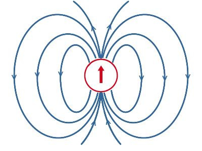
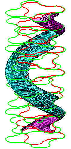
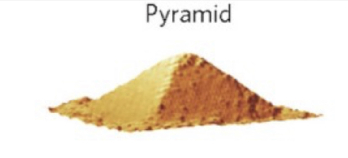
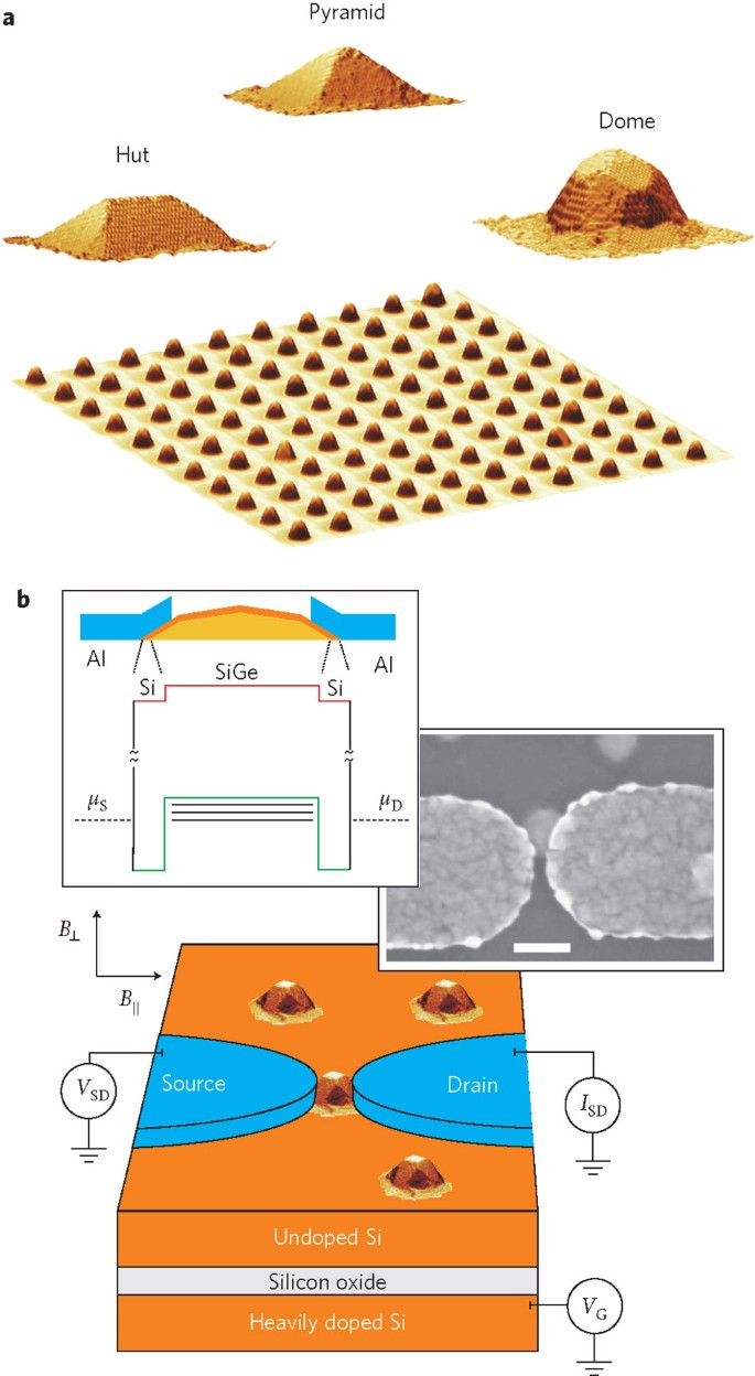
Magnetotactic bacteria usually mineralize either iron oxide magnetosomes, which contain crystals of magnetite (Fe3O4), or iron sulfide magnetosomes, which contain crystals of greigite (Fe3S4). Several other iron sulfide minerals have also been identified in iron sulfide magnetosomes—including mackinawite (tetragonal FeS) and a cubic FeS—which are thought to be precursors of Fe3S4. One type of magnetotactic bacterium present at the oxic-anoxic transition zone (OATZ) of the southern basin of the Pettaquamscutt River Estuary, Narragansett, Rhode Island, United States is known to produce both iron oxide and iron sulfide magnetosomes.
The lipid coating is what they are utilizing for everything!
The word liposome derives from two Greek words: lipo ("fat") and soma ("body"); it is so named because its composition is primarily of phospholipid. Liposomes were first described by British haematologist Alec Douglas Bangham in 1961 (published 1964), at the Babraham Institute, in Cambridge.
Everything is connected! ❤️
🙂


Liposome-quantum dot hybrids: lipid bilayer-embedded hydrophobic QD vesicles loaded with doxorubicin. (A) Schematic depiction of hybrids; (B) cryo-transmission electron microscopy; (C) atomic force microscopy (3D images) of empty liposomes, L-QD hybrids, and L-QD-Dox hybrids (left to right). (D) Serum stability of L-QD-Dox hybrids incubated in 50% mouse serum. QD were embedded in EPC:Chol:DSPE-PEG2000 and DSPC:Chol:DSPE-PEG2000 liposomes. Dox was loaded using the pH-gradient technique and Dox release was assessed by measuring Dox fluorescence. (E) Cytotoxicity of L-QD-Dox hybrids. MCF-7 cells were incubated with free Dox, L-QD-Dox, and L-QD hybrids, and cell viability was assessed using MTT assay. Reprinted with permission from [103], W. T. Al-Jamal and K. Kostarelos, Liposomes: From a clinically established drug delivery system to a nanoparticle platform for theranostic nanomedicine.
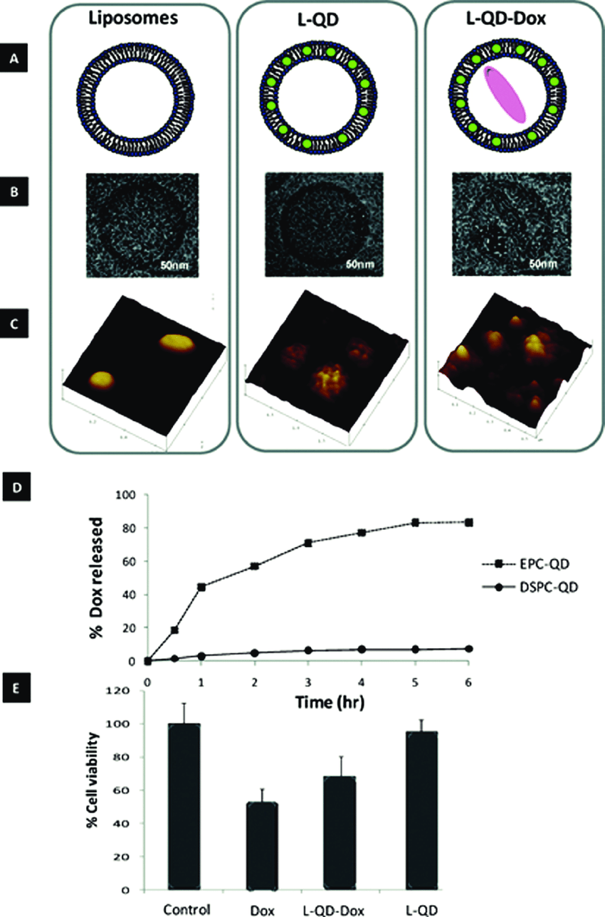
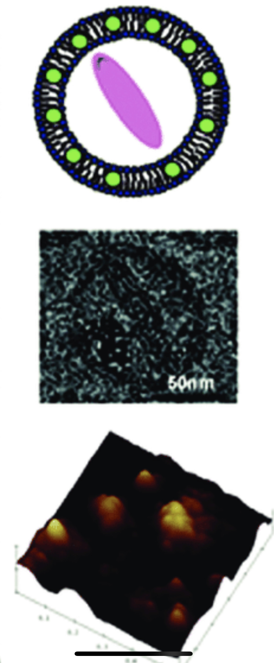


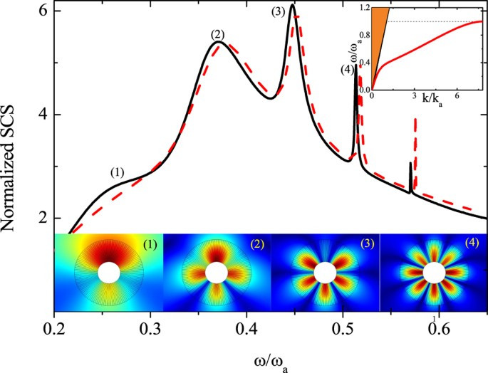
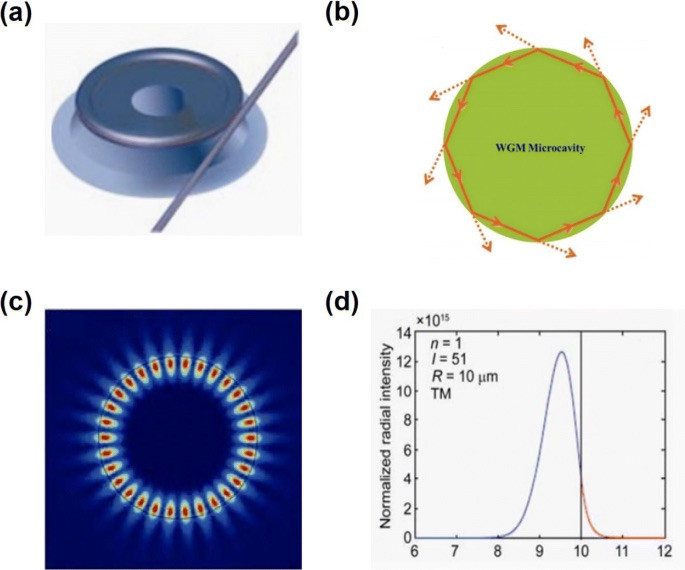
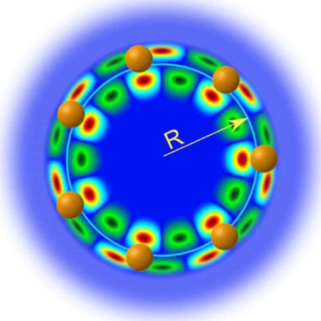
FIG. 1. Illustration of a WGM cavity excited by frustrated total internal reflection at a prism surface. The initial excitation is in the clockwise (CW) direction. Addition of plasmonic gold nanoparticles introduces coupling into the counterclockwise (CCW) direction, resulting in standing wave modes (SWMs) with non-degenerate eigen frequencies. Small biomolecules interacting with the gold surface introduce shifts in the WGM resonance frequency and change the frequency splitting of the SWMs

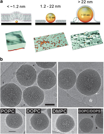
Supported lipid bilayers (SLBs) on nanotopographic solid substrates. (a) Top: schemes illustrate the structures of lipid bilayers in the presence of silica nanoparticles with varying size.
Bottom: corresponding atomic force microscopy (AFM) images of upper schemes.16 (b) Top: cryo-transmission electron microscope images from mixtures of silica particles and 1,2-dioleoyl-sn-glycero-3-phosphocholine (DOPC)/1,2-dioleoyl-sn-glycero-3-phospho-L-serine (DOPS) (4:1) small unilamellar vesicles (SUVs), quickly frozen after a 1-min incubation time. Most particles present one or two patches of lipid bilayer at their interfaces (black arrows). Few particles present the adsorbed, unruptured vesicles at their surface (white asterisks). Bottom: SLBs formed with SUVs containing neutral lipids only (DOPC, 1-palmitoyl-2-oleoyl-sn-glycero-3-phosphocholine (POPC) and L-α-dimyristoyl phosphatidylcholine (DMPC)) and a high concentration of negatively charged lipids (DOPC/DOPS, 1:1). The scale bars are 50 nm.18
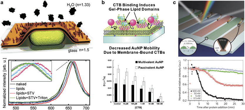
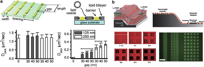
Lipid-nanostructure hybrids for lipid membrane modulation. (a) Top: schematics of a substrate with intermittent nanogaps (left) and cross-sectional view of a supported lipid bilayer (SLB) formed on a nanopatterned substrate. Bottom: diffusion coefficients of lipids in a perpendicular direction to metallic nanogap patterns (right) were significantly modulated compared with those in a parallel direction (left).77 (b) Top: SLB on a topographic wall creating elastic energy barriers at both side ends (white lines). Bottom left: time evolution of gel phase domain formation in an arrayed nanosmooth region. The epifluorescence signal comes from Texas-red 1,2-dihexadecanoyl-sn-glycero-3-phosphoethanolamine (DHPE). The scale bar is 200 μm. Bottom right: an epifluorescence microscope image of gel phase microdomain arrays reacted with Alexa 488-CTB (cholera toxin subunit B). The scale bar is 200 μm.82
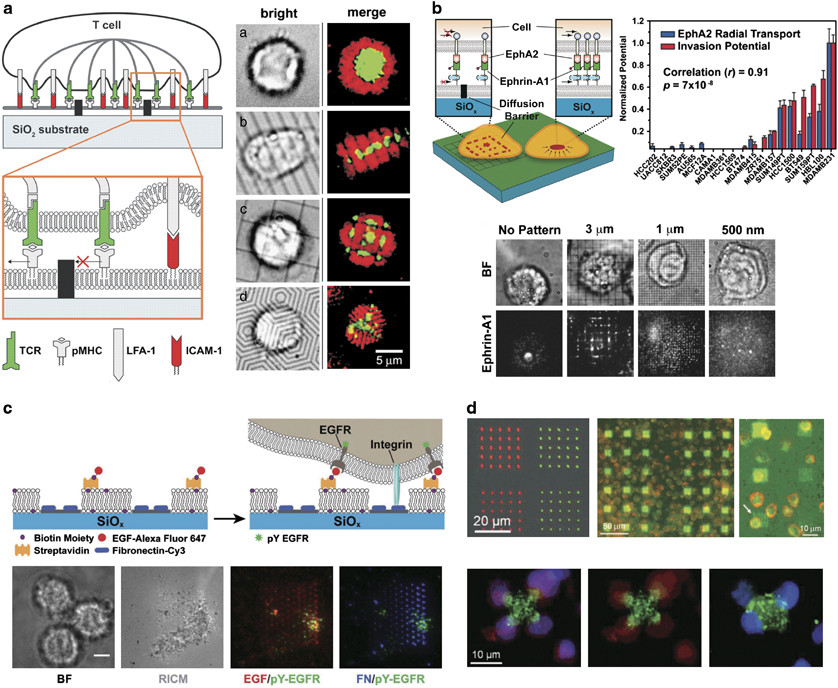
Interfacing synthetic lipid bilayers with live cells. (a) Left: schematic diagram of a live T-cell-supported membrane junction. Right: bright field and fluorescence images of the T cell and ligands. Synapse formation is altered by the geometrical constraints of the substrate. T-cell receptor (TCR; green) and intercellular adhesion molecule 1 (ICAM-1) (red) were labeled with fluorescent tags.63 (b) Left: experimental supported lipid bilayer (SLB) platform that triggers and manipulates the EphA2 receptors on the surface of living cells.
Right: average ephrin-A1 ligand radial distribution functions for 26 cell lines are quantified and parameterized. The average radial distribution function was found to exhibit a strong correlation (r=0.91, P=7 × 10⁻⁸) with invasion potentials that were determined with the modified Boyden chamber analysis. Bottom: images of fluorescently labeled ephrin-A1 ligands of EphA2-expressing mammary epithelial cells on a supported membrane.87 (c) Schematic illustration of the binary ligand system that presents immobilized FN-Cy3 and laterally mobile EGF-647. Representative brightfield, RICM, and fluorescence microscopy images of three cells that are engaged with a nanopatterned supported membrane.32 (d)
The fluorescence image of phospholipid nanopatterns deposited on a glass surface. A three-channel image shows the T-cells adhering to the corners of lipid-protein dip-pen nanolithography (DPN) patterns and activated by anti-CD3/anti-CD28 (green=lipid pattern, blue=the nucleus of cells by 4,6-diamidino-2-phenylindole (DAPI), red=CD69 by anti-CD69-phosphatidylethanolamine (PE) and anti-PE-tetramethylrhodamine-5(and-6)-isothiocyanate (TRITC).30
This study also showed the importance of the lipid composition in constructing a continuous and intact lipid bilayer. Complete SLB formation on a silica nanoparticle was achieved using liposomes with positive, neutral or low net negative charges, whereas liposomes with a high net negative charge remained unruptured on silica nanoparticles (Figure 1b). The size of the small unilamellar vesicles also has a profound effect on the kinetics of the vesicle-to-bilayer transformation on nanostructures pitted with nanoholes. ...
nanohole (plural nanoholes)
(physics) A hole of nanoscale dimensions; but especially one of an array of such holes on a semiconductor surface.
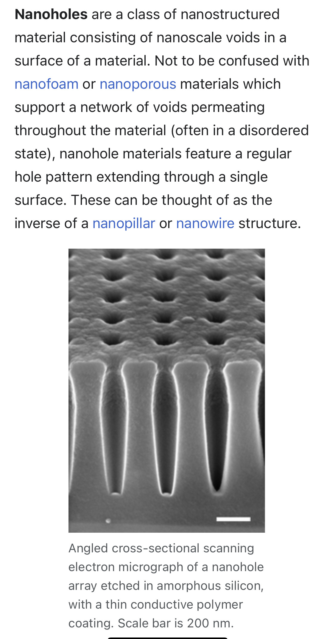
Nanohole structures have been used for a variety of applications, ranging from superlenses produced from a metal nanohole array,[1] to structured photovoltaic devices used to improve carrier extraction,[2] and light absorption.[3]
Nanohole structures are also extensively utilized for the creation of photonic crystals, particularly for creating photonic crystal waveguides.