So basically they (the satanic peodophiles) have manipulated you with nanoparticles disguised as a virus.
... vacccine???
Are peodophiles the direct result of vaccines that cause damage to the brain?
🤒It is also caused by the substances in plastics and in the medicines. Allready 20 year ago there was a study that proved that some medicines (like Prednison) caused lesbianity in woman. And weak makers in plastics are causing lower male hormones, resulting in more gay men. 'These people are sick' > Q is right: they are poisoned and became sick. The clowns who did this on purpose are evil.
Are you trying to tell me that vaccines cause oxidative stress that leads to brain trauma injury that leads to the onset of peodophillia?
Surely this is immoral?
Agreed.
@themac
Question: do you know what is Luciferase? Can it be that it is jelly-fish nematocystic electrolyt with activ ions in it ? That would explains the persons with burning wounds after the vaxx. A side effect of this electrolyt could be that it is working/attracks Cu ions: so that it is working on ceruloplasmine, a Cu enzym for the making of globuline (a substance in the bloodplasma)? If so, what could work against it to neutralize this luciferase substance?
(Why am I thinking this about luciferase: because [we] are researching a weird case on very old jelly-fishes that are petrifyed in a weird way: they are saved as bournonite like metal disks? And they differ from pyriet in staying perfect yellow without oxydising. Could it be that the nematocystic ions attract metal ions that preserved them? )
This could explain the bloodcloths in the vaxxed?
Luciferase-displaying protein nanoparticles were genetically designed for use as highly sensitive detection probes.
Certain nanoparticle probes possess several unique properties that are advantageous for use in the detection of cancer at the early stages. In this review, we will discuss the advances in the development of nanoparticle-based methods for the detection of cancer by fluorescence spectroscopy.
Nano probes are used for focusing on imaging, visualization and detection of diseases. ... Such Nano probes exhibit tunable optical, spectroscopic, magnetic, and electrical properties for signal amplifications.
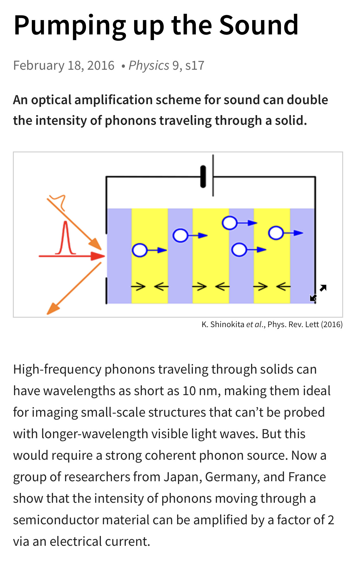
Acoustic phonons are the main heat carriers in graphene near room temperature while optical phonons are used for counting the number of atomic planes in Raman experiments with few-layer graphene.
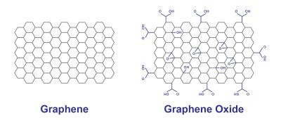
Graphene oxide is an oxidized form of graphene, laced with oxygen-containing groups. It is considered easy to process since it is dispersible in water (and other solvents), and it can even be used to make graphene. Graphene oxide is not a good conductor, but processes exist to augment its properties.
In this view, we recently demonstrated the ability of a few layer graphene (FLG) and different GOs to induce a sustained cytotoxicity related to a reduced mitochondrial activity in human skin HaCaT keratinocytes, a preliminary in vitro model to investigate skin toxicity.8 It is widely known that one of the main causes of mitochondrial dysfunction is represented by oxidative stress.9 Intriguingly, it has been suggested that oxidative stress plays a major role in the cytotoxicity of other carbon-based nanomaterials.10 The link between reactive oxygen species (ROS) overproduction and mitochondrial damage is well known. Doubtlessly, mitochondria are both the source and target of ROS, the latter being able to induce an oxidative stress-caused mitochondrial damage. Therefore, it is crucial to shed light on the role of ROS in the mechanism of cytotoxicity induced by GBNs.
graphene-based nanomaterials (GBNs)
Coating graphene oxide nanoflakes with cationic lipids leads to highly homogeneous nanoparticles (GOCL NPs) with optimised physicochemical properties for gene delivery applications. In view of in vivo applications, here we use dynamic light scattering, micro-electrophoresis and one-dimensional sodium dodecyl sulfate polyacrylamide gel electrophoresis to explore the bionano interactions between GOCL/DNA complexes (hereafter referred to as "grapholipoplexes") and human plasma. When exposed to increasing protein concentrations, grapholipoplexes get covered by a protein corona that evolves with protein concentration, leading to biocoronated complexes with modified physicochemical properties.
Here, we show that the formation of a protein corona dramatically changes the interactions of grapholipoplexes with four cancer cell lines: two breast cancer cell lines (MDA-MB and MCF-7 cells), a malignant glioma cell line (U-87 MG) and an epithelial colorectal adenocarcinoma cell line (CACO-2).
Luciferase assay clearly indicates a monotonous reduction of the transfection efficiency of biocoronated grapholipoplexes as a function of protein concentration. Finally, we report evidence that a protein corona formed at high protein concentrations (as those present in in vivo studies) promotes a higher capture of biocoronated grapholipoplexes within degradative intracellular compartments (e.g., lysosomes), with respect to their pristine counterparts. On the other hand, coronas formed at low protein concentrations (human plasma = 2.5%) lead to high transfection efficiency with no appreciable cytotoxicity. We conclude with a critical assessment of relevant perspectives for the development of novel biocoronated gene delivery systems.
Keywords: bionano interactions; cationic lipids; gene delivery; graphene oxide; protein corona.
Gene delivery is the process of introducing foreign genetic material, such as DNA or RNA, into host cells. Gene delivery must reach the genome of the host cell to induce gene expression.
In optogenetic systems, engineered photoreceptor proteins are used to transduce a light stimulus, ultimately resulting in enhanced or reduced gene expression [12], [13]. The photoreceptors are typically bound to a chromophore, a small molecule ligand that undergoes a chemical change upon light absorption.
The production of new proteins in response to external stimuli results largely from rapid activation of gene transcription — this is known as inducible gene expression. ... Inducible genes are highly regulated and must be able to be rapidly and specifically activated in response to stimuli.
A destabilized version of firefly luciferase was used in living yeast cells as a real-time reporter for gene expression. This highly sensitive and non-invasive system can be simultaneously used upon many different experimental conditions in small culture aliquots. This allows the dose-response behaviour of gene expression driven by any yeast promoter to be reported and can be used to quantify important parameters, such as the threshold, sensitivity, response time, maximal activity and synthesis rate for a given stimulus.
We applied the luciferase assay to the nutrient-regulated GAL1 promoter and the stress-responsive GRE2 promoter. We find that luciferase expression driven by the GAL1 promoter responds dynamically to growing galactose concentrations, with increasing synthesis rates determined by the light increment in the initial linear phase of activation. In the case of the GRE2 promoter, we demonstrate that the very short-lived version of luciferase used here is an excellent tool to quantitatively describe transient transcriptional activation.
The luciferase expression controlled by the GRE2 promoter responds dynamically to a gradual increase of osmotic or oxidative stress stimuli, which is mainly based on the progressive increase of the time the promoter remains active. Finally, we determined the dose-response behaviour of a single transcription factor binding site in a synthetic promoter context, using the stress response element (STRE) as an example. Taken together, the luciferase assay described here is an attractive tool to rapidly and precisely determine and compare kinetic parameters of gene expression.
The brain in conscious control of genes—the idea sounds more outlandish than it should. After all, brain-computer interfaces, wireless power, and optogenetics already exist. It’s just that nobody thought to combine them before. Correcting this oversight, researchers at ETH Zurich assembled a system that has allowed humans to control gene expression by doing nothing more than adopting different mental states, or thinking different thoughts.
Thus far, the system has been used to control gene expression only in cell cultures and mice. But the system’s creators are hopeful that mind-controlled gene switches, which represent the integration of cybernetics and synthetic biology, could be refined to control gene expression in people.
Recent advances in optical technologies such as multi-photon microscopy and optogenetics have revolutionized our ability to record and manipulate neuronal activity. Combining optical techniques with electrical recordings is of critical importance to connect the large body of neuroscience knowledge obtained from animal models to human studies mainly relying on electrophysiological recordings of brain-scale activity. However, integration of optical modalities with electrical recordings is challenging due to generation of light-induced artifacts.
Here we report a transparent graphene microelectrode technology that eliminates light-induced artifacts to enable crosstalk-free integration of 2-photon microscopy, optogenetic stimulation, and cortical recordings in the same in vivo experiment. We achieve fabrication of crack- and residue-free graphene electrode surfaces yielding high optical transmittance for 2-photon imaging down to ~ 1 mm below the cortical surface. Transparent graphene microelectrode technology offers a practical pathway to investigate neuronal activity over multiple spatial scales extending from single neurons to large neuronal populations.
As an intriguing two dimensional material, graphene has attracted intense interest due to its high stability, large carrier mobility as well as the excellent conductivity. The addition of graphene into the heterogeneous electrodes has been proved to be an effective method to improve the energy storage performance.
Graphene electrodes could enable higher quality imaging of brain cell activity thanks to new research by a team of engineers and neuroscientists at the University of California San Diego.
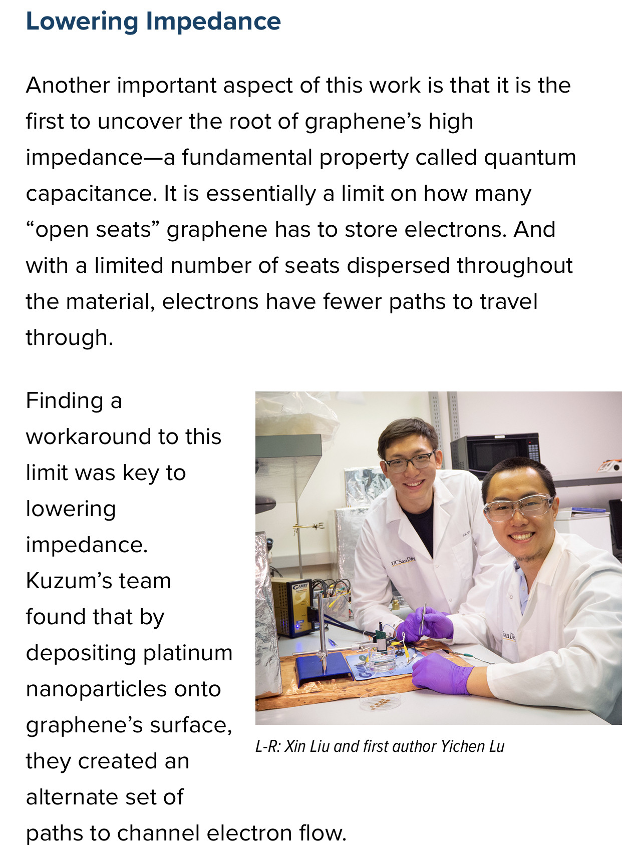
The researchers developed a technique, using platinum nanoparticles, to lower the impedance of graphene electrodes by 100 times while keeping them transparent. In tests on transgenic mice, the low-impedance graphene electrodes were able to record and image neuronal activity, such as calcium ion spikes, at both the macroscale and single cell levels. The advance brings graphene electrodes a step closer to being adapted into next-generation brain imaging technologies and various basic neuroscience and medical applications.


