nagative lipid + positive lipid
Dipole, literally, means "two poles," two electrical charges, one negative and one positive.
We say that neurons have an electrochemical charge, and this charge changes, depending on whether the neuron is at rest or is sending a signal.
An electric dipole deals with the separation of the positive and negative charges found in any electromagnetic system. A simple example of this system is a pair of electric charges of equal magnitude but opposite sign separated by some typically small distance. (A permanent electric dipole is called an electret.)
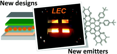
It is inevitable that electroluminescent devices, notably light-emitting diodes (LEDs), organic LEDs (OLEDs), and light-emitting electrochemical cells (LECs), will heat up during operation because of Joule heating,[1] the formation of dark and quenched excitons,[2] and self-absorption.[3] This self-heating is an unfortunate and problematic issue, since a too high operational temperature results in a severe drop of the device efficiency and premature device failure.[4] Accordingly, a significant number of studies on the cause and consequences of self-heating, and on how to alleviate its effects, are available in the scientific literature for both LEDs[1] and OLEDs.[1, 4, 5]
A LED is a compact point emitter that features a very high luminance from a small spot; it is, as such, clearly different from an OLED or a LEC, which are thin-film surface emitter
Thin-film electrodes (TFEs), which consist of a layer of active material with a thickness ranging from nanometers to micrometers, have been widely explored in the field of supercapacitors (SCs)—especially thin-film SCs, flexible and/or stretchable SCs, and microsupercapacitors (MSCs).
https://pubs.acs.org/doi/10.1021/acsnano.1c04996#
Lipid Nanoparticles—From Liposomes to mRNA Vaccine Delivery, a Landscape of Research Diversity and Advancement
Rumiana Tenchov, Robert Bird, Allison E. Curtze, and Qiongqiong Zhou*
Surface charge is a key determinant of nanoparticle fate in vivo1,2. Following intravenous (i.v.) injection, nanoparticles with high surface charge density, either anionic or cationic, are rapidly cleared from circulation by specialised cells of the reticulo-endothelial system (RES)3,4,5. In mammals, RES cell types are primarily located in the liver (key hepatic RES cell types: Kupffer cells, KCs, and liver sinusoidal endothelial cells, LSECs) and spleen.
These cells are responsible for clearing up to 99% of i.v. administered nanoparticles from circulation6. High nanoparticle surface charge density has a qualitative and quantitative impact on serum protein binding7,8,9,10,11,12, driving the opsonisation of circulating nanoparticles and subsequent recognition and clearance by the RES13,14,15. In addition, cationic nanoparticles tend to adsorb to the anionic surface of cells and are subsequently internalised16,17,18,19,20, often leading to acute cytotoxicity21,22,23. Given the adverse pharmacokinetics of charged nanoparticles in the body, most clinically approved, nanoparticle-drug formulations (nanomedicines) possess a (near) neutral surface charge to prolong circulation lifetimes and maximise drug exposure within target (vascularized) tissues in the body24
A series of thin InN films down to 10 nm in thickness were prepared by molecular-beam epitaxy on either AlN or GaN buffers under optimized growth conditions. By extrapolating the fitted curve of sheet carrier density versus film thickness to zero film thickness, a strong excess sheet charge was derived, which must come from either the surface or the interface between InN and its buffer layer. Since metal contacts, including Ti, Al, Ni, and a Hg probe, can always form an ohmic contact on InN without any annealing, it is determined that at least part of the excess charge is surface charge, which was also confirmed by capacitance–voltage measurements. © 2003 American Institute of Physics.


